Skin Cancer FAQs
Basal cell carcinoma, squamous cell carcinoma, and melanoma are the most common and well-known skin cancers and arise from the most common cell types in the skin. However, a variety of less common cell types found in the skin can also become malignant and lead to more rare cancers such as atypical fibroxanthoma/undifferentiated pleomorphic sarcoma, Merkel cell carcinoma, mycosis fungoides/cutaneous T cell lymphoma, dermatofibrosarcoma protuberans, extramammary Paget’s disease, Kaposi’s sarcoma, microcystic adnexal carcinoma, and others. There are over seventy-five different types of skin cancer and most of them are very rare and named to make doctors feel like they got their money’s worth out of medical school (e.g. endocrine mucin-producing sweat gland carcinoma, extranodal NK/T-cell lymphoma nasal type, primary cutaneous CD8+ aggressive epidermotropic cytotoxic T-cell lymphoma, primary signet ring carcinoma of the eyelid, spindle cell liposarcoma, etc.).
Skin cancer is the most common kind of cancer in the United States, which makes sense given that it is also the largest organ in the human body. More than 3 million people are diagnosed with more than 5 million skin cancers annually. Although many skin cancers occur on sun-exposed skin in fair-skinned people, they can occur anywhere on the body and in any skin tone.
UV-exposed skin is more likely to develop skin cancers because the ultraviolet (UV) radiation from the sun damages DNA, and mutated DNA is more likely grow out of control and become malignant (cancerous). Sunburns from childhood and young adulthood seem to have the greatest impact on DNA. However, the more overall UV radiation the skin has to handle, the more likely you are to develop a skin cancer. Cumulative UV exposure does matter.
UV radiation damage to DNA persists throughout life, even if it happened years ago. We don’t appreciate the damage until we get older because DNA repair machinery generally corrects the damage. And if the repair machinery misses something, the immune system identifies and destroys it. However, the machinery and the immune system wear out with time, just like everything else in the body, and become less efficient. We start to see the damage that doesn’t get corrected and that can lead to cancer. And the more UV radiation that the skin has to process on top of historical damage, the greater the risk for developing skin cancer. Cumulative exposure is the primary risk factor for nonmelanoma skin cancers (BCC and SCC).
Although genetic and environmental factors may be contributing factors, cancers likely develop in sun protected areas because the immune system’s surveillance missed a bad cell that happened to develop while normal cells were dividing. Once a cancer cell escapes detection, it has the opportunity to establish itself and grow into a tumor.
Skin cancer can be found in people of all ages and ethnicities. However, it is most common in people with fair complexions, red or blond hair, and blue eyes. Genetics also plays a role. There are several genetic conditions associated with skin cancer.
Skin Cancer Prevention
There are many possibilities to choose from to help protect your skin. Not every option is going to be the answer for every person. However, it does take a little personal commitment and perhaps a mindset adjustment to see the opportunities for self-care rather than the burden of behavioral change.
- Use a facial lotion with an SPF of 30 or higher year-round
- Avoid tanning bed (spray tans are great!)
- Wear protective SPF clothing if planning to be outdoors for an extended amount of time (then you don’t have to remember to re-apply the sunscreen!)
- Perform self-skin exams and look for new or changing moles: take monthly photos with your phone for comparison.
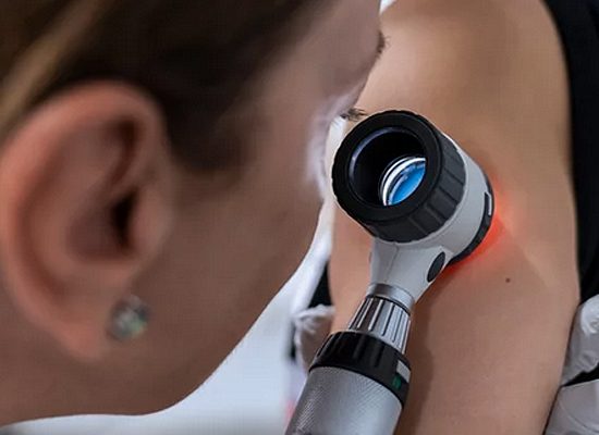
- See your dermatologist yearly for a full body skin exam.
- If you’re worried about it, get it checked.
- Wear a hat with a brim when outdoors: you want to protect your ears, nose, and mouth. Ball caps only protect the scalp and forehead, which are probably the least cosmetically important places on the head and neck.
Types of Skin Cancer
Squamous cell carcinoma (SCC) is the second most common kind of skin cancer and develops from the squamous cells that make up the middle and outer part of the epidermis. Sometimes dermatologists will refer to this type of cancer as cutaneous SCC (cSCC) in order to clarify that a skin cancer is being discussed, because SCC also develops in the mouth, throat, lungs, and other locations that have squamous linings.
SCC has a widely variable appearance. It can be a persistent, thick, rough, scaly patch that may crust or bleed. It can resemble a wart that crusts or bleeds. It can be a non-healing open sore, especially if it’s at the edge of an old burn or chronic ulcer. It may look like a little volcano with a central core of keratin. The common thread is that regardless of shape, it doesn’t heal or go away.
Most squamous cell carcinomas of the skin are caused by too much UV radiation, either from the sun or tanning beds. Protecting your skin from UV light can help reduce the risk of squamous cell carcinoma of the skin. SCC is usually not life-threatening but can become dangerous if ignored. While most cutaneous SCCs remain localized and easily treated, roughly 3-5% can become regionally, nodally, or widely metastatic. This most often occurs in immunosuppressed patients, those with long-neglected tumors, and/or those with aggressive tumor subtypes.
Like BCC, the specific treatment options depend on the location, tumor subtype, and size of the SCC. Very superficial SCCs may be treated with electrodessication and curettage, intense liquid nitrogen, chemotherapy cream, or photodynamic therapy. Larger cancers will need electrodessication and curettage, excision with safety margin, Mohs micrographic surgery, intralesional chemotherapy, or radiation therapy. If the cancer has spread into nerves, bone, or even further beyond the skin, then adjuvant radiation, chemotherapy, or immunotherapy may be needed.
Melanoma is the third most common skin cancer and arises from melanocytes. These are the cells that make pigment, called melanin, which gives the skin its color. Melanoma is the most lethal of the common skin cancers, but can be easily treated with an excellent prognosis when it is caught early.
Melanoma typically starts on sun-exposed skin, but it can develop anywhere that has melanocytes. This means that it can occur inside the eyes/mouth/genitals, on the palms/soles, or in the beds of the fingernails/toenails. These so-called “hidden” melanomas are more common in skin of color. The exact cause of all melanomas isn’t clear, but most are caused by exposure to ultraviolet light. Roughly 10% are familial and related to a specific genetic mutation. Limiting exposure to UV light can help reduce the risk of melanoma.
Melanoma can first appear as a change in an existing mole or, more commonly (about 70 to 80% of the time), as the development of a growth on seemingly normal skin. The ABCDEs (described below) for evaluating pigmented lesions are very helpful in identifying potential melanomas. Looking for the “Ugly Duckling” mole can also be useful because moles are usually similar on a given body. If one mole looks substantially different from the rest, it might be because it is a cancer. Unfortunately, not all melanomas are brown. Some are “amelanotic”, which means that they lack pigment and therefore may be pinkish, reddish, white, skin-colored, or even clear and colorless. This makes them hard to identify, even for dermatologists. And this is why knowing your own skin is so critical. You are more likely to notice a new skin-colored spot than your doctor is and can monitor it with photos to determine if it changes over time. If it changes, get it checked.
ABCDE’s of Melanoma
- A- Asymmetry: One half of the mole is unlike the other side
- B- Border: Irregular or poorly defined border
- C- Color: Varying colors throughout the mole
- D- Diameter: Usually greater than 6 millimeters (about 0.24 in), or about the size of a pencil eraser
- E- Evolving: Mole is changing (can be size, shape, or color)
Melanoma treatment depends on the depth of the cancer at diagnosis, referred to as the Breslow thickness, certain tumor characteristics, and the localization of the cancer. Most melanomas are found early enough that they are treatable with only a surgical excision that removes any visible remaining tumor with a safety margin of normal-appearing skin. If the Breslow thickness is beyond a certain depth, then an excision and a sentinel lymph node biopsy are usually done at the same time. A sentinel lymph node biopsy is a procedure that determines whether or not the melanoma cells have spread to a nearby lymph node, which is usually the first place that melanoma cells travel to when they metastasize. Further treatment depends on complete staging work-up, which may include MRI, PET scan, and/or CT. Immunotherapy, targeted therapy, chemotherapy, and radiation are all options for treating advanced melanoma. This treatment is managed by an oncologist and radiation oncologist. The five-year relative survival rate for localized (skin only) melanoma is >99%. It drops to 74% for regional melanoma and 35% for distant melanoma.
Mohs Surgery for Skin Cancer
Mohs surgery involves removing a cancer layer by layer and looking at each layer under the microscope until no cancer cells are left. This allows the surgeon to remove the whole growth without taking too much of the healthy skin around it. It is followed by reconstruction of the defect with plastic surgery techniques. Mohs surgery has the highest cure rate of any method to treat squamous cell and basal cell carcinoma. It is not used to treat invasive melanoma.
Other Treatment Options
- Electrodessication and Curettage (ED&C) This treatment involves scraping away the skin cancer with a rounded blade called a curette (curettage). Cancer has a different consistency when scraping, similar to a bruise within an apple, that lets the physician feel when they’ve reached healthy skin. Then an electric needle is used to sear the edges of the cancer and destroy any residual malignant cells (electrodessication). The curette is then used to remove this tissue. Several rounds of this are done to make sure all of the cancer is destroyed.
- Freezing. This treatment, called cryosurgery, involves freezing cancer cells with liquid nitrogen. This is much more intense than the freezing used to treat an actinic or seborrheic keratosis. It requires anesthesia of the area because the lesion has to undergo two cycles at a temperature of -50°C in order to kill the tumor cells. Sometimes the freezing is done after using a scraping tool, called a curette, to remove the surface of the skin cancer.
- Photodynamic therapy. During photodynamic therapy, a liquid medicine that makes the cancer cells sensitive to light is applied to the skin. After this is absorbed by the damaged skin, a light that destroys the skin cancer cells is shined on the area to activate the medication. The light is usually blue in color and thus this treatment is also referred to as “Blue Light Therapy”. This treatment might be used with surgery or other treatments.
- Excision. This involves cutting out the lesion in an ellipse shape with a margin of healthy skin around it. The amount of tissue removed depends on the specific lesion being excised. Benign lesions require margins of 2-3 mm all the way around whereas SCCs, BCCs, and melanoma in-situs (MIS) need 5 mm margins. Invasive melanoma may require a 10 mm or 20 mm margin depending on the depth. Sometimes the excision is done at the same time that a lymph node receiving drainage from the cancerous area is sampled to see if the cancer has spread. This manner of testing the draining lymph node is called a sentinel lymph node biopsy.
- Radiation therapy. Radiation therapy uses powerful energy beams to kill cancer cells. Radiation therapy is sometimes used after surgery when there is an increased risk that the cancer might return. This is called adjuvant radiation. It also might be an option for people who can’t have or don’t want surgery. Radiation for skin cancer has a cure rate ~90%, the treated area cannot be re-treated with radiation in the future, and the irradiated skin is paradoxically prone to skin cancer development after many years. For this reason, it is not a good choice for younger patients. Much lower doses of radiation are also used after keloid excisions because the radiation greatly increases the success rate of treatment.
- Intralesional chemotherapy. Injection of chemotherapy directly into the lesion can be an effective treatment for certain types of SCC. 3-4 injections may be needed over the course of several weeks. The cure rate is less effective than surgery but this can be a nice alternative to surgery for treating multiple small SCCs.
- Chemotherapy. Chemotherapy uses medicine that disrupts cell growth to kill cancer cells. Chemotherapy can be used alone or with other treatments, such as targeted therapy, immunotherapy, and radiation therapy.
- Targeted therapy. Targeted therapy uses medicines that attack specific chemicals in cancer cells. By blocking these chemicals, targeted treatments eliminate cancer cells. Targeted therapy is usually used with chemotherapy.
- Immunotherapy. Immunotherapy is a treatment with medicine that helps the body’s immune system kill cancer cells, which often survive by hiding from the immune system. Immunotherapy helps the immune system find the cancer cells and destroy them.
Not all treatment options will be advisable for all types of skin cancer. In many cases, more than one treatment would be reasonable to treat a given malignancy. In these instances, you and your dermatologist should discuss the risks and benefits for each option and together choose the method that best fits your particular cancer and circumstances.